**Discovery of Vacuoles:**
– Contractile vacuoles first observed by Spallanzani in protozoa.
– Dujardin named vacuoles in 1841.
– Schleiden applied the term for plant cells in 1842.
– De Vries named the vacuole membrane tonoplast in 1885.
**Functions of Vacuoles:**
– Isolate harmful materials.
– Contain waste products.
– Maintain internal hydrostatic pressure.
– Maintain acidic internal pH.
– Assist in autophagy and lysis of misfolded proteins.
**Types of Vacuoles:**
– Central vacuoles: found in most plant cells, surrounded by tonoplast membrane, maintain turgor pressure, store chemicals for cell protection.
– Contractile vacuoles: specialized osmoregulatory organelles in protists, regulate water and ion balance.
**Vacuoles in Pathology:**
– Vacuolization as an unspecific sign of disease.
– Need for more research to establish links between vacuolization and specific diseases.
– Vacuolization observed in various biological specimens.
– Implications of vacuolar dysfunction in diseases like Parkinson’s.
– Research ongoing to identify therapeutic targets related to vacuoles.
**Cell Biology of Vacuoles:**
– Membrane-bound organelles in cell cytoplasm.
– Store substances like water, nutrients, and waste products.
– Large central vacuole in plant cells maintains turgor pressure.
– Regulate cellular processes and maintain cell structure.
– Variability in size and function of vacuoles across different cell types.
A vacuole (/ˈvækjuːoʊl/) is a membrane-bound organelle which is present in plant and fungal cells and some protist, animal, and bacterial cells. Vacuoles are essentially enclosed compartments which are filled with water containing inorganic and organic molecules including enzymes in solution, though in certain cases they may contain solids which have been engulfed. Vacuoles are formed by the fusion of multiple membrane vesicles and are effectively just larger forms of these. The organelle has no basic shape or size; its structure varies according to the requirements of the cell.
| Cell biology | |
|---|---|
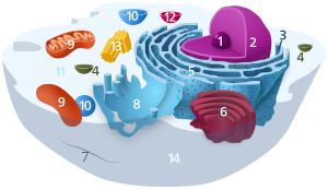
| Animal cell diagram | |
 Components of a typical animal cell:
|


